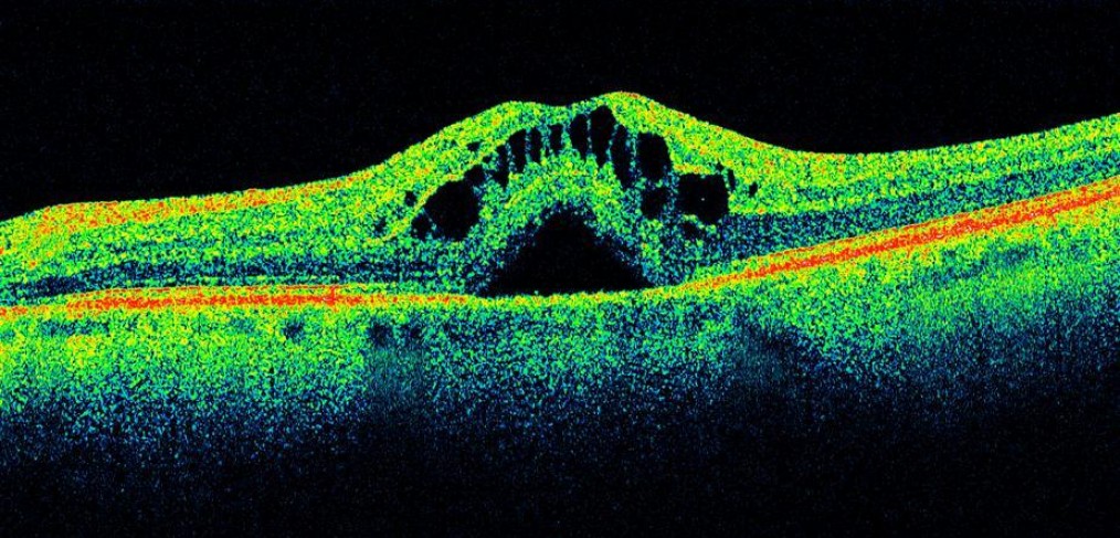
OCT – Optical Coherence Tomography
Optical coherence tomography (OCT) is a recent non-invasive imaging technique that performs a “luminous CT scan,” which allows to analyse the retinal structures with high resolution sections of the retina. The new generation OCT Spectral Domain has a resolution of about 4 micron, which allows an almost histological reconstruction of retinal tissue. Moreover, the acquisition of each optic tomography has a duration of a fraction of a second and is highly reproducible.
The scans obtained can then be analysed, quantified, protected, compared with subsequent scans and superimposed to fluorangiographic images, ICGA and microperimetric maps. OCT Spectral Domain can be replaced by fluorangiography in a certain number of conditions, avoiding the patient the need to submit to an invasive examination. The high resolution of OCT Spectral Domain allows the doctor to define the functional meaning of the retinal structures to better understand the diseases. Briefly, OCT Spectral Domain is a reliable, sensitive, reproducible, non-invasive examination that allows to study the retina layer be layer, almost histologically, obtaining “optic biopsies” and also identifying the finest possible lesions that cause retinal diseases.
OCT is useful in the diagnosis, stadiation and follow-up of many retinal diseases, including maculopathies, hereditary retinal dystrophies, macular oedema in diabetic retinopathy or in vascular venous occlusions, central serous chorioretinopathy and diseases of the vitreous-retinal interface.
