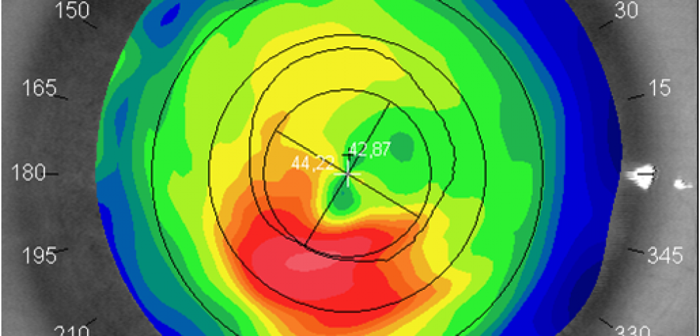
TOPO – Corneal topography
Corneal topography is a non-invasive examination that consists of photographic acquisition of the corneal surface. This technology is irreplaceable in the study (diagnosis, follow-up, pre and post-operative path) of many corneal diseases, refractive surgery, contactology in cases of high astigmatism or warpage caused by contact lenses, and in all cases of irregular surfaces. Besides said indications, corneal topography is used to screen and follow-up keratoconus, a particular progressive corneal astigmatism that gradual exhausts the cornea. If it is not treated early, it can lead the patient to corneal transplantation.
The evolution of topographs (in which the industries, technicians and Italian ophthalmologists have played a leading role) and, subsequently, the considerable increase in information that can be obtained, has made it quite complex to interpret maps; hence the need, in the first place, to clearly know what to highlight in a printed report of the disease of the examined patient.
It is important to keep in mind that, in the field of ophthalmology, unlike geographic-cartography, conventional topography does not draw the shape of the cornea but represents it in map form with encoded colours (hence it would be more precise to speak of photokeratoscopy or of computerised video keratoscopy). It is obtained by adding the points on the anterior corneal surface, covered by tear film, on which the curvature radius is calculated and, subsequently, the refractive power.
